

 |
 |
 |
FT-IR microscopy is a very powerful technique to characterize the chemical composition of natural and synthetic fibers.
Introduction
The identification of fibers is of high importance in forensic science as it can yield trace evidence in a criminal case. A fiber found at the crime scene can provide information about the criminal, e.g. about his clothing or specific places he has visited. Furthermore the fiber might be compared to similar items retrieved from suspects and provide the link to the crime. In textile industry the identification of individual fibers in the textile matrix is interesting as a part of the quality control process, e.g. to track an unwanted polymer fiber used during the spinning process of a yarn. Also for conservators of textile artifacts the ability to differentiate between fibers originating from different natural materials (e.g. cotton, sisal, bast, silk and wool) is very helpful to choose an appropriate treatment.
FT-IR microscopy is a very powerful technique to characterize the chemical composition of natural and synthetic fibers, both organic and inorganic in nature. Due to its capability to measure with a high lateral resolution usually the availability of a single fiber is sufficient to perform an analysis. As the FT-IR measurement is non-destructive other analytical techniques might be applied afterwards. Furthermore, FT-IR microscopy provides objective results and is in most cases quicker, easier, and sometimes, more selective than classical methods. Due to these multiple benefits the IR-microscopic method is described by ASTM International as standard method for forensic analysis of fibers (ASTM E2224-10) and for identification of fibers in textiles (ASTM D276-12). Using the ATR- (Attenuated Total Reflectance) technique minimal sample preparation is required to perform an FTIR- microscopic measurement. Just a fixation of the fiber on a flat substrate like a metal plate is required to avoid its movement during visual inspection and definition of the measurement positions. In this application note measurements of different natural and synthetic fibers using the fully automated FT-IR microscope LUMOS are presented.
Instrumentation
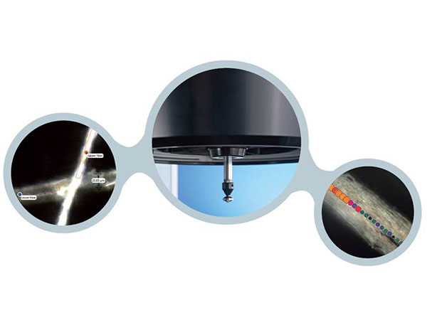
The presented fiber measurements were performed using the stand-alone FT-IR microscope LUMOS (Fig.1). It stands out due its full automation and ease-of-use combined with sample visualization and infrared spectroscopic performance of excellent quality. Its 8x objective provides the measurement modes ATR, transmission and reflection and high quality visual inspection capabilities. All required changes of hardware settings as well as the complete IR-measurement procedures including background measurements are performed fully automated – even in the ATR-mode which is the typical approach to measure fibers. To provide perfect contact to samples ranging from soft to very hard the ATR-device offers three pressure steps and is equipped with a very precise internal pressure sensor.
Due to a large working distance and an unobstructed access to the planar sample stage the sample positioning is extremely convenient. Additionally for maximum performance and convenience the LUMOS includes:
• Motorized Germanium ATR crystal with internal pressure control
• Large field of view: 1.5 x 1.2 mm
• Automated change of the numerical aperture between IR and Vis mode to achieve a high depth of field for the isual inspection of a sample, but also highest sensitivity for the IR analysis
• Independent white light LED illumination in transmission and reflection
• Fast CMOS camera with 4x zoom
• Motorized stage (option), position accuracy of 0.1 μm
• Optional macro accessory that allows to use all Quick
• Snap sampling modules from the compact FTIR spectrometer ALPHA
Example: Chemical Imaging of a human hair IR-microscopy even allows detecting and visualizing chemical variances on the surface of a single fiber. This example shows the measurement of a human hair that has been partially bleached. To expand the area on which the mapping measurement can be performed and to prevent sample deformation due to the contact with the ATR-crystal the hair was flattened using a diamond compression cell prior to the measurement. A line map was performed along the hair covering a bleached and a non-bleached (outgrown) part. The bleaching affects the fingerprinting range of the IR-spectra, e.g. bands at 1180cm-1 and 1040cm-1 increase in intensity. Figure 7 shows the chemical image based on the integration of the 1040cm-1 band superimposed with the visual image of the hair. With the integration intensities at each measurement position being color and size coded the image clearly shows the chemical difference between bleached and unbleached hair.
Summary
IR-microscopy is an established technique to determine the chemical identity of synthetic and natural fibers. The imaging capabilities of the IR-microscopic method even allow visualizing chemical differences on single fibers with high lateral resolution. With the fully automated standalone IR-microscope LUMOS the analysis of fibers can be performed without specific IR-spectroscopic expertise. The intuitive software guided workflow allows even untrained personal quickly performing the measurement and spectra evaluation.
Credit : Bruker Co., Ltd.


|
|
|