

 |
 |
 |
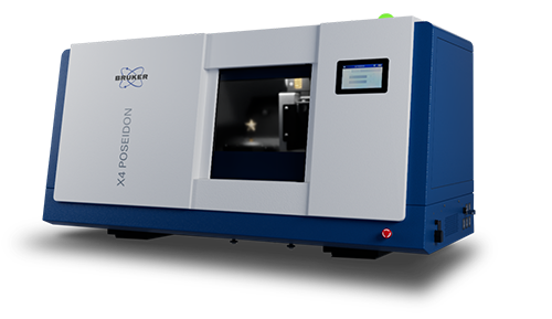
The X4 POSEIDON is a powerful micro-computed tomography machine that delivers the functionality of a floor-standing system in a compact benchtop design.
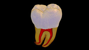
Micro-CT, also known as 3D X-ray microscopy (3D XRM), is an advanced imaging technique that provides an in-depth 3D view of samples for biomedical, life and materials science.
The X4 POSEIDON is a modular X-ray tabletop microscope. The instrument can be upgraded according to changing needs and, as the technology evolves, new components can be easily integrated.
The X4 POSEIDON is a comprehensive micro-CT solution from the start featuring many premium features to enhance the user experience.
In its essential configuration the X4 POSEIDON includes a proven reflection X-ray source and versatile flat panel detector. With these components, the system is capable of ultra-fast scanning with exceptional field-of-view and razor-sharp contrast without compromising resolution.
Whether your lab supports multiple users in diverse research and development fields or a specific industrial process, the X4 POSEIDON offers the full power of nondestructive, qualitative and quantitative 3D analysis.
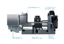
By combining Best-Scan-Geometry and Multi-Vision, the X4 POSEIDON delivers ultimate flexibility in a benchtop with Geometric Magnification Plus - GEM Plus™.
A balance of field-of-view and resolution is achieved by moving the sample closer to the source or closer to the detector. With Best-Scan-Geometry, the detector can also be moved in a continuous way, allowing ideal cone beam utilization for a given sample size while achieving the same magnification ratio, resulting in a significant scan speed improvement.
GEM Plus™ builds on Multi-Vision and Best-Scan-Geometry to strike the perfect balance between pure resolution, scan speed and field-of-view.
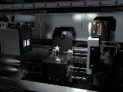
The X4 POSEIDON is the only true multi-detector benchtop XRM solution. It can be equipped with a versatile flat-panel detector, a scientific-grade CMOS detector, or both!
When selecting the ideal detector for your needs, you must consider the balance between field-of-view, resolution and scan speed. If speed is of upmost importance, the flat-panel detector is ideally suited for your application. On the other hand, the sCMOS detector is the ideal choice for maximum resolution.


|
|
|