Ultimate Ease-of-Use
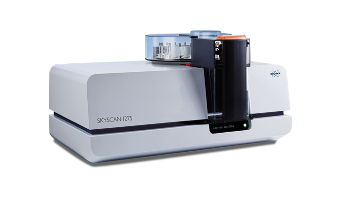
The desktop SKYSCAN 1275 is a real 3D X-ray microscopy power pack, designed for fast scanning of a wide range of samples. Thanks to the compact geometry and fast flat-panel detector the time to result can be as short as a few minutes. Just ideal for high throughput imaging.
The SKYSCAN 1275 comes furthermore with a high level of automation thus providing superior ease-of-use. Simply pushing the button at the control panel starts an auto-sequence of a fast scan, followed by reconstruction and volume rendering. All executed while the next sample is already being scanned. The optional 16-position sample changer even allows for unattended high throughput scanning.
The SKYSCAN 1275 is complemented by 3D.SUITE. This extensive software suite covers GPU-accelerated reconstruction, 2D/ 3D morphological analysis, as well as surface and volume rendering visualization.
Key Features
Push-Button XRM
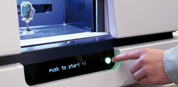
Just insert a sample, manually or automatically, and get a complete 3D volume without any further interaction.
Push-Button XRM includes everything: automatic sample size detection, sample scanning, 3D reconstruction, and 3D volume rendering. Ideal for routine tasks, or operation by non-expert users!
You need full control? No problem, SKYSCAN 1275 offers all features.
Combined with a sample changer SKYSCAN 1275 even works 24/7.
Sample changer
Ex vivo scanning of biological tissues is a very good way to show their internal structures non-destructively. A contrast agent or chemical drying can improve the image quality by further enhancing or differentiating densities. The SKYSCAN 1272 is the ideal platform for such imaging with its resolution, easy sample handling and high throughput – that’s why you will find so many publications using this scanner.
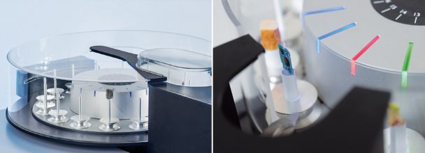
SKYSCAN 1275 can optionally be equipped with an external 16-position sample changer to increase throughput for QC and routine analysis.
The sample changer accepts a variety of sample sizes, up to a diameter of 96 mm.
Samples can be can be easily replaced at any time without interrupting an ongoing scanning process. New samples are automatically detected, and LED‘s indicate the status for every scan: ready, scanning, done.
In-situ stages
MicroCT is exceptionally good for the visualization of internal structures in the finest details of the tissues of plants and animals. This imaging method differentiates between densities without harming or destroying the scanned object. That’s why zoology and botany are fast-growing microCT applications with SKYSCAN 1272 users at the forefront. A wide range of different living organisms can be visualized and analyzed with minimal or no sample-treatment.
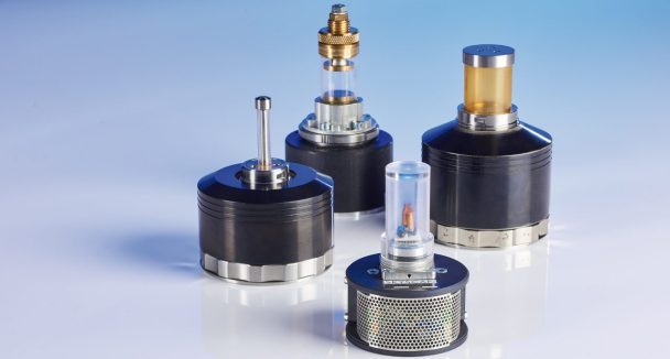
The Bruker material testing stages are designed to perform compression experiments up to 4400 N and tensile experiments up to 440 N. All stages automatically communicate through the system’s rotation stage, without the need of any cable connections. Using the supplied software, scheduled scanning experiments can be set up.
Bruker's heating and cooling stages can reach temperatures of up to +80ºC, or 30ºC below ambient temperature. Just like the other stages, no extra connections are needed, and there is an automatic recognition of the stage. Using the heating & cooling stages, samples can be examined under non-ambient conditions, to evaluate the effect of temperature on the sample’s microstructure.
Highlighted Applications
Bone Applications
The SKYSCAN 1275 provides the bone research community with a solid solution for fast, high throughput and high-quality bone morphometry of animal models. The advanced cMOS flat panel camera cuts rodent morphometry scans to a tiny fraction of what scientists are used to with the first generation of osteoporosis research microCT scanners – we’re talking about 5 minutes only. Without compromise to resolution of the smallest rod-like mouse trabecular structures – nothing is lost. With the SKYSCAN 1275 you will publish more papers each year than with scan-slow microCT alternatives.
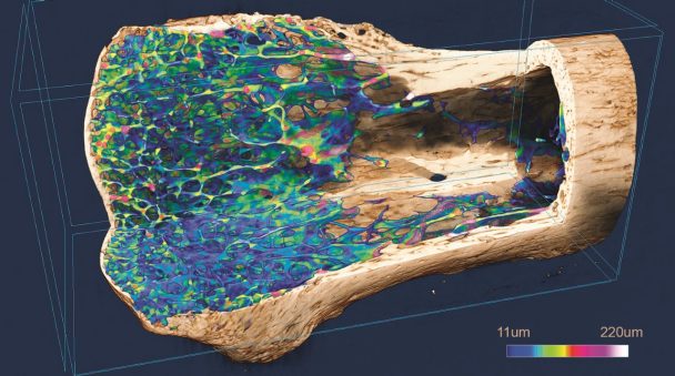
- Fast cMOS flat panel with ~2000 x 1500 pixel format, fastest scans less than 100 seconds (step and shoot).
- Effortless imaging of large bone samples not previously studied by microCT with fast throughput for research productivity.
- All rodent morphometry of trabecular and cortical bone is taken care of, with bone morphometry (ASBMR nomenclature) with comprehensive 3D and 2D parameters, and densitometry including BMD calibration references over a 2-32mm diameter size range.
- Optional sample changer for 16 preloaded samples makes large scale screening studies possible with thousands of mouse or zebrafish bones for trabecular and cortical bone parameters.
Dentistry
Teeth are a step up from rodent bones in thickness and mineral density, requiring an appropriate microCT solution. The SKYSCAN 1275 is this solution, effortlessly imaging small and large teeth, even fossil ones subject to mineral diagenesis. And this capability in a user-friendly high-throughput desktop instrument. With the SKYSCAN 1275 you will publish more papers each year than with scan-slow uCT alternatives.
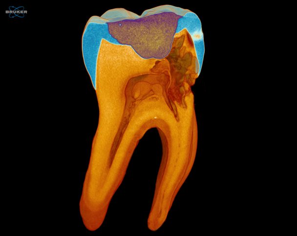
- Fast cMOS flat panel with ~2000 x 1500 pixel format, fastest scans less than 100 seconds (step and shoot).
- The large x-ray camera with excellent high energy sensitivity makes most vertebrate teeth scannable as well as prostheses and dental implant materials.
- Optional sample changer for 16 preloaded samples makes large scale dental screening and endodontic before-and-after canal treatment studies straightforward.
- Bone morphometry (ASBMR nomenclature) with comprehensive 3D and 2D parameters, with densitometry including BMD calibration references over a 2-32mm diameter size range.
Plant and Animal Biology
MicroCT is exceptionally good at visualization of the finest details of the internal structure of biological tissue. It is already a well-established tool in botany and zoology. The SKYSCAN 1275 is a unique compact and user-friendly desktop scanner with a powerful flat panel camera allowing – in a nutshell – faster scanning of bigger samples than before. Its fast, user-friendly scanning with a large field of view removes barriers to the advantageous use of this non-destructive 3D imaging technique to more and more scientists and disciplines.
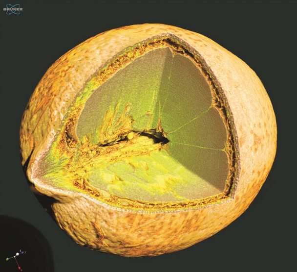
- Fast evaluation of the internal structures of fruits, seeds, insects, corals and marine organisms.
- Digitize a 3D volume of valuable samples for further visualization or cataloging.
- Multiple sample scanning possible due to the batch-scanning mode and optional sample changer, effectively reducing down-time.
- Comprehensive 3D image analysis capability including morphometry and densitometry, 3D registration, segmentation and advanced image processing methods.


