Advance Analytical
เครื่องมือวิเคราะห์ทางวิทยาศาสตร์ขั้นสูง Advance Analytical
Advance Analytical
Hits: 4455

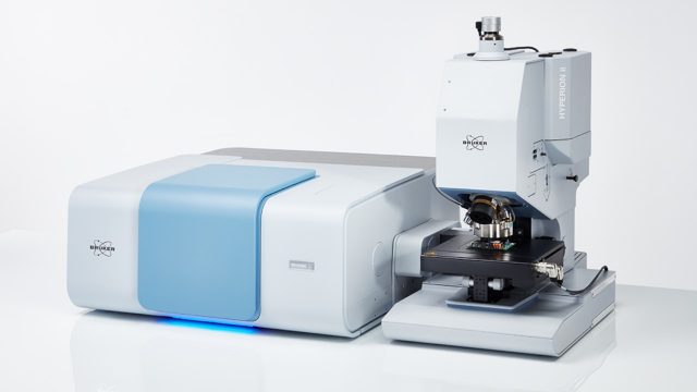
The HYPERION II is an innovation force in infrared microscopy. It provides IR imaging down to the diffraction limit and sets the benchmark in ATR microscopy. It combines FT-IR and Infrared Laser Imaging (ILIM) microscopy for the first time ever in a single device, offering all three measurement modes: transmission, reflection, and ATR.
HYPERION II features:
- Selection of detectors for µ-FT-IR: Broad-, mid, narrow-band LN2-MCTs,thermoelectrically cooled (TE) MCT.
- Focal-plane array detector for infrared imaging (64 x 64 or 128 x 128 pixel).
- Optional QCL implementation by Laser Infrared Imaging Module (ILIM, laser class 1)
- Objective lens selection: 3.5x/15x/36x IR, 20x ATR, 15x GIR, 4x/40x VIS.
- Spectral range extension – from Near-Infrared (NIR) to Far-Infrared (FIR)Selection of apertures: manual knife-edge, automated knife-edge aperture wheel. Metal apertures for NIR
- Selection of accessories and sample stages: macro IR imaging accessory, cooling/heating stage, sample compartment, etc.
- Selection of visual/optical tools: Darkfield illumination, Fluorescence illumination, VIS polarizers, IR polarizers, etc.
HYPERION II provides:
- Perfect match of spectral and visual images. Applies to any measurement mode (including ATR imaging).
- Diffraction limited high sensitivity FT-IR microscopy and imaging by using focal plane array (FPA) detector
- First ever combination of FT-IR and QCL technology by (optional) Infrared Laser Imaging Module (ILIM, laser class 1).
- Infrared laser imaging in all measurement modes (ATR, Transmission, Reflectance).
- Patented coherence reduction for artifact free Laser Imaging measurements without sensitivity or speed loss.
- High imaging speeds: 0.1 mm2per second (FPA, full spectrum) 6.4 mm2 per second (ILIM, single wavenumber)
- Optional TE-MCT detector to perform IR microscopy with high spatial resolution and sensitivity without liquid Nitrogen.
- Emission spectroscopy capability and optional spectral range extensions.
HYPERION II applications:
- Life science | cell imaging
- Pharmaceuticals
- Emissivity studies (e.g. LEDs)
- Failure and Root Cause Analysis
- Forensics
- Microplastics
- Industrial R&D
- Polymers & Plastics
- Surface characterization
- Semiconductor

Reliable Identification of Microplastics of any Dimension, on any Filter
Microplastic particles (MPP) are by definition polymer particles ranging in size from 5.000 to 1µm. They originate from polymer beads added to cosmetic and personal care products as well as abrasion of macroscopic objects such as plastic bottles, synthetic textiles, and car tires.

Fast Imaging of Large Biological Tissue Samples
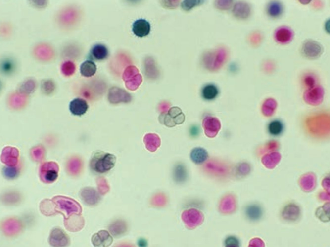
FTIR microscopy has developed into a powerful tool for the analysis of biological tissue. The obtained IR spectral images allow for the differentiation of components and the identification of malignant or diseased tissue.

Defect Analysis using FT-IR Microscopy
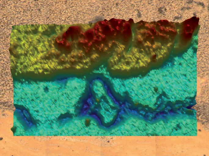
Microscopic techniques are suitable for locating and analyzing defects as small as a few millimeters or even micrometers for many different types of products. Fourier-Transform infrared (FT-IR) microscopy is capable of revealing the chemical composition of such failures with a high spatial resolution.

Analysis of composite films
FT-IR microscopy is a very effective method for the analysis of composite films. Using FT-IR microscopy allows identifying both material and thickness of the individual layers in a foil.

Infrared Microspectroscopic Imaging of Biological Samples
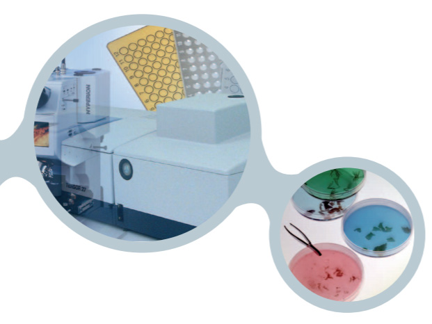
FT-IR imaging is a powerful technique to generate chemical images showing the distribution of the main biochemical components (e.g., lipids, proteins, and polysaccharides) in plant and animal tissue. Moreover, the obtained spectral information allows to differentiate between various properties
of the analyzed tissue, including healthy and diseased.

New & exciting application fields open up to the IR spectroscopy
(FT-) IR spectroscopy has become an indispensable tool in the chemical and pharmaceutical industry
for characterizing materials and identifying substances.Pharmaceutical and biotechnological companies use this technique to analyze both proteins and cellular systems in order to develop new drugs and products.
Contact us
Syntech Innovation Co., Ltd.
388/5 Nuanchan Road, Nuanchan,
Buengkum, Bangkok 10230
388/5 Nuanchan Road, Nuanchan,
Buengkum, Bangkok 10230
0 2363 8585 (auto)
0 2363 8595
081 498 9939

3089208
Today
Yesterday
This Month
All days
166
1549
28916
3089208
Your IP: 216.73.216.80
2026-02-17 02:25


