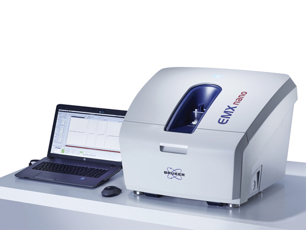
microESR is a small, portable research grade instrumentThe microESR is a small, portable research grade instrument. The spectrometer has a mass of only 10 kg and a 30.5 x 30.5 x 30.5 cm3 foot print. It can easily fit in a fume hood or glove box, or be transported to the field. It requires no special installation or regular maintenance.
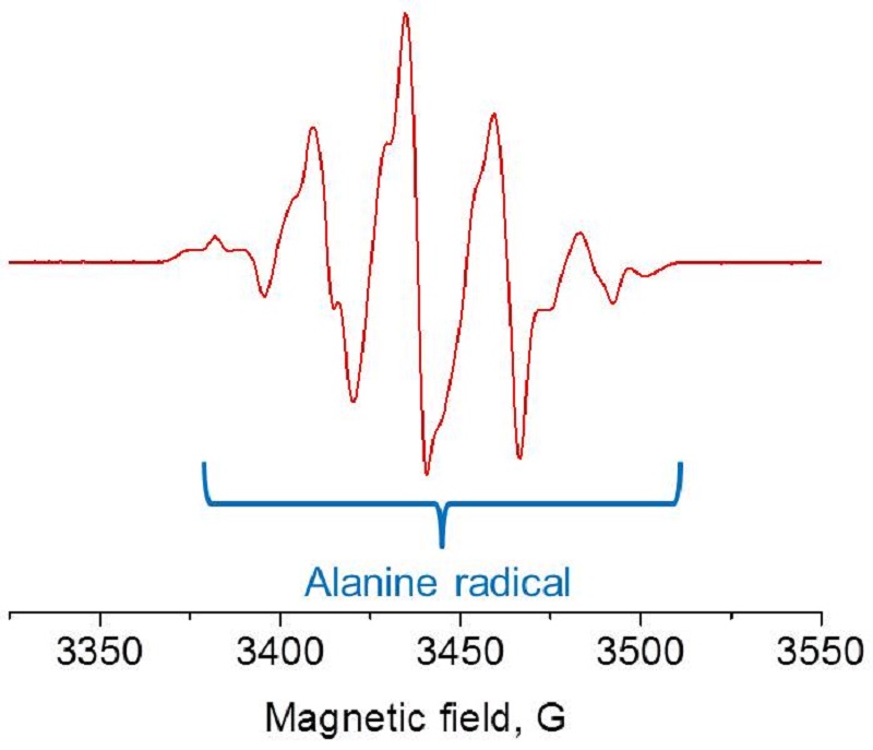
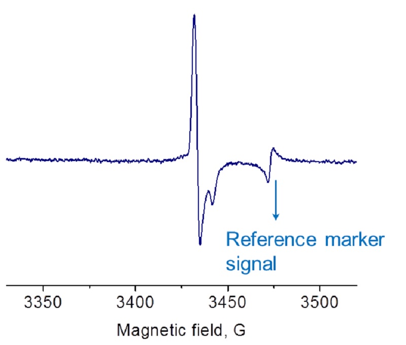
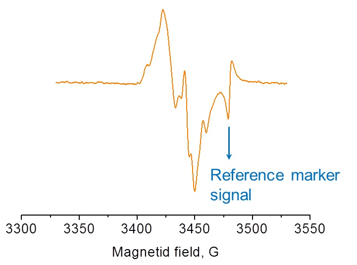
The microESR is also an ideal teaching tool for undergraduate chemistry labs. This instrument enables classroom demonstrations of both simple topics such as free radicals in everyday life to far less intuitive subjects including electron density, spin-orbit coupling, spin-spin exchange, and forbidden transitions. The Education Package is a very good investment for Chemistry Departments as there are a wide range of labs and subjects that can be addressed with the microESR.
- Operating Frequency: X-Band
- Continuous Wave
- Field Sweep Range: 500 G centered at g=2
- Spectrum simulation and fitting
- Easily run samples at liquid nitrogen temperature
ApplicationEducationResearch Grade Teaching Tool
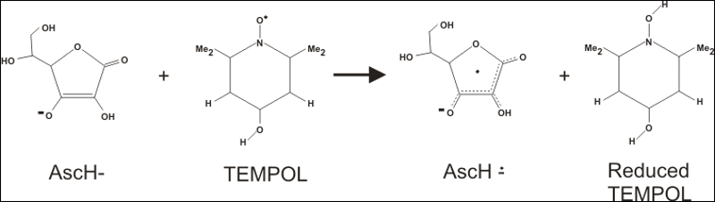
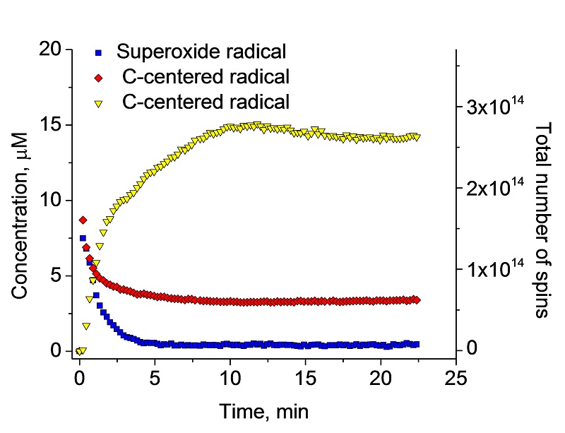
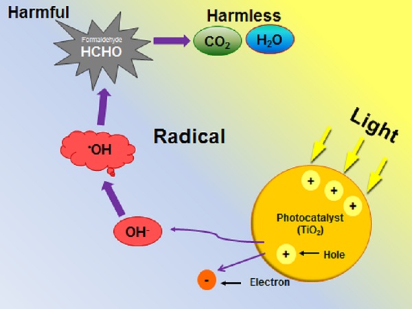
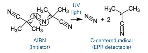
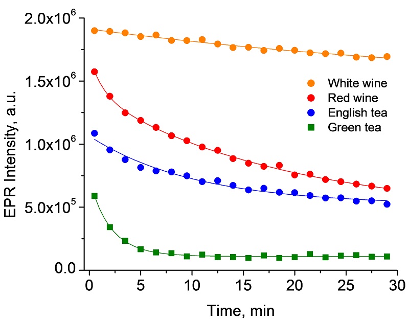
ChemistryReaction kinetics, free radical chemistry, catalysts, DNP
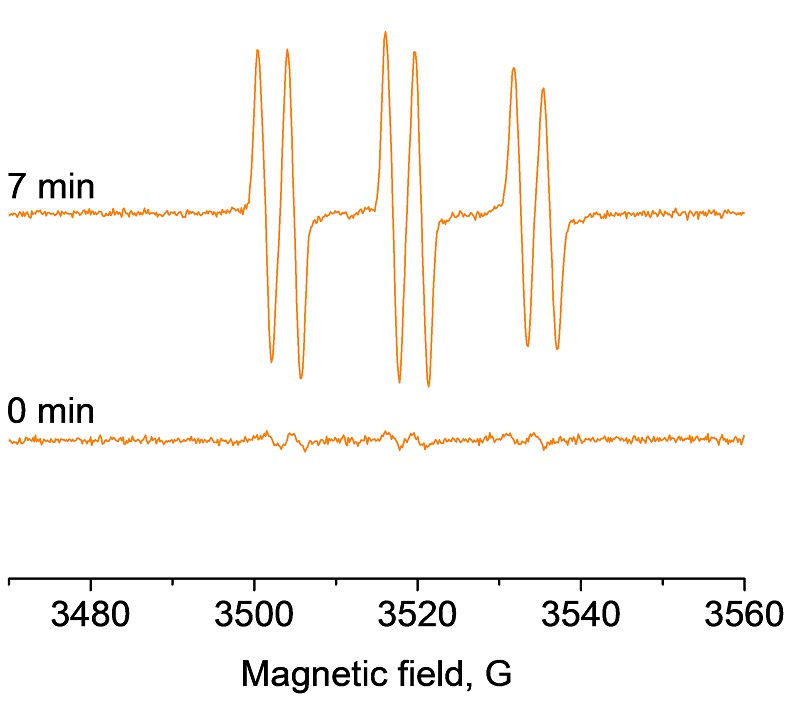
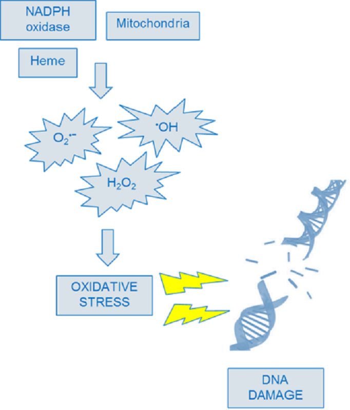
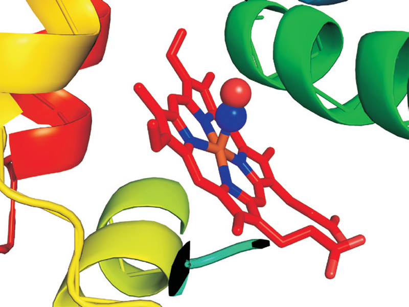
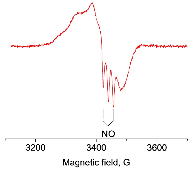
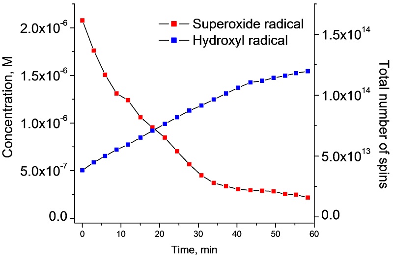
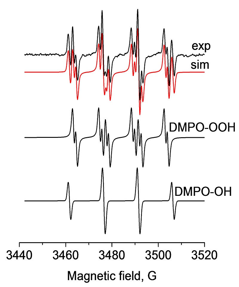
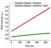
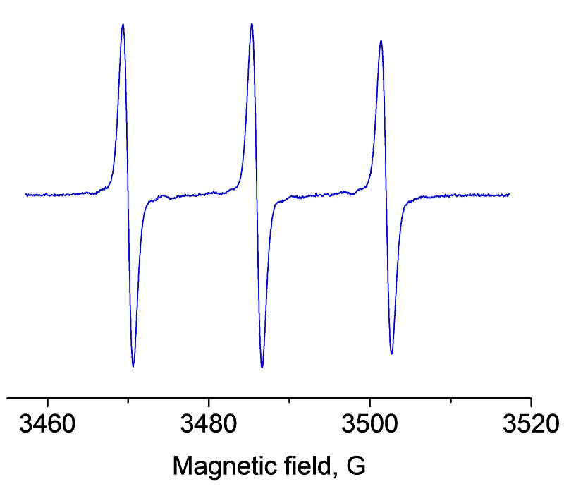
Biology/Biochemistry/BiomedicalSpin labeling, spin trapping, nitric oxides, ROS and RNS
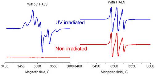
Materials SciencePolymer degradation
IndustryFree radicals in polymers and polymerization, petrochemistry, thermoxidative breakdown of lubricants and fuel, real time analysis of additives, antioxidants in lubricants and fuels.